
Fig. S7
- ID
- ZDB-FIG-170206-13
- Publication
- Stratman et al., 2017 - Mural-Endothelial cell-cell interactions stabilize the developing zebrafish dorsal aorta
- Other Figures
- All Figure Page
- Back to All Figure Page
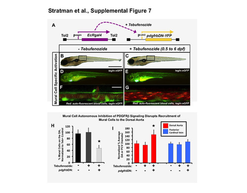
Mural cell-autonomous inhibition of pdgfr signaling leads to loss of mural cell coverage on the zebrafish dorsal aorta. (A) Schematic diagram showing the Tg(tagln:EcRgal4) and Tg(uas:pdgfrbDN-YFP) transgenes used for heat shock-inducible expression of dominant negative PDGF receptor beta (pdgfrbDN). These fish were outcrossed to Tg(tagln:egfp) transgenic fish to generate triple heterozygotes, treated with tebufenozide for the indicated time frame, and imaged for analysis of mural cell coverage at 6 dpf. (B-G) Representative 6dpf images of untreated (B,D,F) or 0.5-6 dpf tebufenozide-treated (C,E,G) Tg(tagln:egfp), Tg(tagln:EcRgal4), Tg(uas:pdgfrbDN-YFP) triple-transgenic animals. Images show whole-animal transmitted light images (B,C), whole-animal green epifluorescence images of tagln:egfp green fluorescence (D,E), and higher-magnification confocal images of the dorsal aorta, showing green fluorescent EGFP-positive vSMC and red autofluorescent circulating red blood cells marking the vascular compartment (F,G). (H) Quantification of the percent number of mural cells covering the dorsal aorta with or without tebufenozide treatment and activation of dominant-negative pdgfrbDN. Fish were treated with the chemical as described above, imaged at 6 dpf and the number of tagln/sm22+ cells per three-somite segment length of dorsal aorta counted. The data represent the % number of tagln/sm22+ cells normalized to the control tebufenozide-treated condition (second column). n= 5 fish from a single experiment; 2 experimental replicates, 2 independent fish clutches per experiment. In more absolute values, we typically see approximately 20 ± s.e.m vSMCs recruit to a 3 somite segment of dorsal aorta under control conditions, and approximately 9-10 ± s.e.m vSMCs recruit to a 3 somite length of dorsal aorta under “DN” activation conditions. (I) Quantification of dorsal aorta (left columns, red) and posterior cardinal vein (right columns, blue) diameter, demonstrating markedly larger dorsal aorta in tebufenozide-treated Tg(tagln:egfp), Tg(tagln:EcRgal4), UAS:pdgfrbDN-YFP) triple- transgenic animals expressing pdgfrbDN autonomously in mural cells. No significant effects were noted on the diameter of the cardinal vein. 3-5 fish were measured per sample, with 10 separate dorsal aorta measurements per fish; experiment repeated twice with consistent results. Values were normalized to the tagln/sm22+ cell counts from non- tebufenozide-treated control fish (column 1). Mean ± s.e.m.; * = p ≤ 0.05 significance from control. Scale bars = 1 mm for B,C and 50 μm for F,G. |