
Fig. S5
- ID
- ZDB-FIG-170206-11
- Publication
- Stratman et al., 2017 - Mural-Endothelial cell-cell interactions stabilize the developing zebrafish dorsal aorta
- Other Figures
- All Figure Page
- Back to All Figure Page
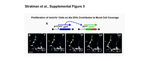
twist1+ cells proliferate to contribute to mural cell coverage. (A) Schematic diagram of the transgenes in Tg(Ola-twist:gal4), Tg(uas:kaede) double transgenic fish, in which the Medaka (Oryzias latipes) twist promoter (Ola-‐ twist) is used to drive expression of Gal4 from the Ola-twist:gal4 transgene, which then activates expression of green to red photoconvertible Kaede from the uas:kaede transgene. (B) Selected frames from a confocal time-lapse image series collected from a 2-2.5 dpf Tg(Ola-twist:gal4), Tg(uas:kaede) double-transgenic fish. Two twist1+ ISV- associated cells highlighted in yellow and green undergo cell division over the course of the time-lapse imaging experiment. The image reconstructions shown are lateral views of the mid-trunk, with rostral to the left. Confocal images were acquired every 10 minutes and the displayed stills are shown at approximately 40-minute intervals. Scale bars = 50 μm. Still images are representative of data collected from 3 individual time-lapse experiments. |