
Fig. S5
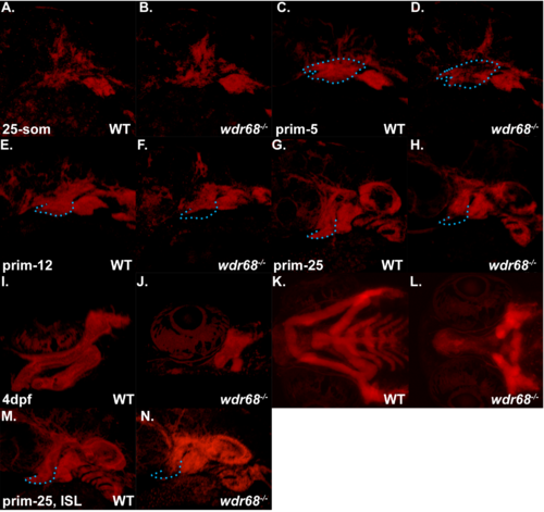
Live confocal analysis of Tg(sox10:mCherryCAAX);wdr68hi3812/hi3812 mutants and wildtype siblings. A-N) Confocal images of the pharyngeal arch regions of embryos live-mounted in 0.7% agarose containing 0.0167% Tricaine. A) 25-somites stage wildtype sibling. B) 25-somites stage wdr68hi3812/hi3812 mutant. C) prim-5 stage wildtype sibling with 1st arch region outlined in blue. D) prim-5 stage wdr68hi3812/hi3812 mutant with same outline as in C to indicate regions of reduced mCherryCAAX signal. E) prim-12 stage wildtype sibling with ventral 1st arch region outlined in blue. F) prim-12 stage wdr68hi3812/hi3812 mutant with same outline as in E to indicate reduced ventral mCherryCAAX signal. G) prim-25 stage wildtype sibling with ventral 1st arch region outlined in blue. H) prim-25 stage wdr68hi3812/hi3812 mutant with same outline as in G to indicate reduced ventral mCherryCAAX signal. I) lateral view of 4-dpf wildtype sibling cartilages. J) lateral view of 4-dpf wdr68hi3812/hi3812 mutant severely reduced M and PQ cartilages. K) ventral view of animal in I. L) ventral view of animal in J. M) ISL-treated prim-25 stage wildtype sibling with ventral 1st arch region outlined in blue. N) ISL-treated prim-25 stage wdr68hi3812/hi3812 mutant with same outline as in M, H, G to indicate modest rescue of ventral 1st arch mCherryCAAX signal. |