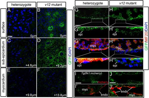
Ventricular expression patterns of the NVclb-EGFP fusion protein. (A,B) GFP expression in the ventricle surface (0 μm) at 52 dpf, showing the formation of plexus-like structures in the v12 mutant (B) compared with heterozygotes (A). (C,D) GFP expression patterns in the sub-epicardium of ventricles at 4.6 μm (C) and 9.2 μm (D) from the surface. Strong expression of GFP fusion protein is shown in the mutant (D) compared with heterozygotes (C). (E,F) GFP is detected at the boundary between endothelial cells and muscles of ventricles at 9.8 μm (E) and 13.8 μm (F) from the surface. (G-H''') GFP is ubiquitously expressed in the ventricle. GFP expression is detected in several layers of GFP+/MF20- epicardium-derived cells in the mutant (H'',H''') compared with a single layer of epicardial cells in heterozygotes (G'',G'''). (I-J′) Tg(flk1:mcherry) transgenic reporter is used to label endothelial cells. GFP is detected at the boundary between endothelial cells (red) and myocardium. endo, endocardium; epi, epicardium; myo, myocardium. Scale bars: 10 μm in A-F,I-J'; 20 μm in G,G'',H,H''.
|
