
FIGURE
Fig. 7
- ID
- ZDB-FIG-160927-25
- Publication
- Jim et al., 2016 - Infection of zebrafish embryos with live fluorescent Streptococcus pneumoniae as a real-time pneumococcal meningitis model
- Other Figures
- All Figure Page
- Back to All Figure Page
Fig. 7
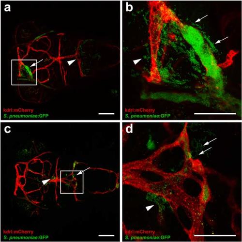
Clogging of the blood vessels by Streptococcus pneumoniae after systemic infection. a-d Confocal microscopy images at maximum projection of Tg(kdrl:mcherry) s896 zebrafish embryos at 4 days post- fertilization injected in the caudal vein. a, c Bacteria were localized inside and outside of the blood vessels with (arrows) and without clogging (arrow heads). Scale bars, 100 µm. b, d An enlarged view of a and c, respectively, with clogging of a blood vessel highlighted. All embryos were infected with 600 CFU and imaged at 24 h post injection. Scale bars, 50 µm |
Expression Data
| Gene: | |
|---|---|
| Fish: | |
| Condition: | |
| Anatomical Terms: | |
| Stage: | Day 4 |
Expression Detail
Antibody Labeling
Phenotype Data
Phenotype Detail
Acknowledgments
This image is the copyrighted work of the attributed author or publisher, and
ZFIN has permission only to display this image to its users.
Additional permissions should be obtained from the applicable author or publisher of the image.
Full text @ J Neuroinflammation