|
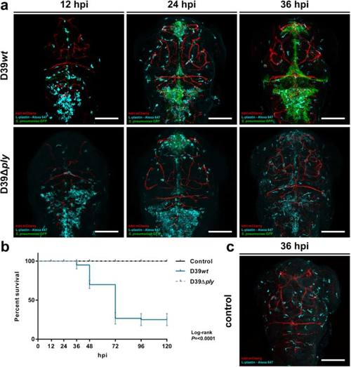
Comparison of 2 days post-fertilization zebrafish embryos infected with wild-type Streptococcus pneumoniae D39 (D39wt) or pneumolysin-deficient mutant strain (D39Δply). a Confocal microscopy images at maximum projection of Tg(kdrl:mcherry) s896 zebrafish embryos infected with D39wt or D39Δply at different time points. c Non-infected zebrafish embryos. D39wt pneumococci grow rapidly compared to D39Δply and migrate throughout the subarachnoid space, delineating the ventricular contours. The numbers of phagocytes reduce over time in the presence of increasing numbers of D39wt bacteria compared to non-infected or D39Δply zebrafish embryos. Embryos were infected with 600 CFU. Scale bars, 100 µm. b Corresponding survival curves. Embryos were infected with 300 CFU. The data represent three individual experiments with 20 embryos in each group
|