Fig. 1
- ID
- ZDB-FIG-160926-9
- Publication
- Thambyrajah et al., 2016 - A gene trap transposon eliminates haematopoietic expression of zebrafish Gfi1aa, but does not interfere with haematopoiesis
- Other Figures
- All Figure Page
- Back to All Figure Page
|
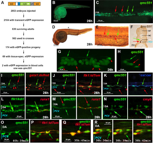
The zebrafish gene trap line qmc551 expresses GFP in primitive red blood cells and in haemogenic endothelial cells of the ventral wall of the dorsal aorta. (A) Structure of the gene trap transposon and strategy of the gene trap screen. (B) Lateral view of a fixed qmc551 embryo. (C) Close-up of the trunk. (D-F) GFP immunohistochemistry and diaminobenzidine staining on a qmc551 embryo. (E) A magnified image of the trunk. (F) A 10 µm transverse section through the trunk of the embryo after plastic embedding. (G) A maximum intensity projection of a 67.5 µm thick confocal Z-stack showing a lateral view of the trunk of a fixed qmc551 embryo. (H) A 1.1 µm YZ cross section of the Z-stack shown in (G). (I,J) Confocal images of the trunk of a qmc551;gata1:dsRed (I) and qmc551;flk1:tdTom (J) double transgenic embryo after fluorescent immunostaining. (K) Confocal images of a live qmc551;csl:cer double transgenic embryo. The csl:cer transgene is a derivative of the csl:venus transgene which we have previously shown to be expressed in arterial ECs ( Gray et al., 2013). (L) Double fluorescent runx1 and flk1/kdrl WISH. (M,N) Fluorescent runx1 (M) and cmyb (N) WISH combined with GFP immunohistochemistry. (O-R) Confocal timelapse microscopy of the DA of qmc551;flk1:tdTom embryos. Images were taken every 3 min. Times on panels represent hours and minutes after fertilization. Note that prRBCs in circulation appear as short lines in the confocal image, while stationary cells are round. The confocal analyses in (I-R) were performed at single cell resolution on 2 (I), 1.8 (J), 2.5 (K), 2.0 (L,N), 1.0 (M) and 2.1 (O-R) µm optical slices. All images (B-E,G,I-R) show embryos with anterior to the left and dorsal up. Red arrows – prRBCs; red arrowheads –- prRBC progenitors trapped in the mesenchyme; green arrows – HECs in the vDA. |
| Genes: | |
|---|---|
| Fish: | |
| Anatomical Terms: | |
| Stage Range: | Prim-5 to Prim-25 |
Reprinted from Developmental Biology, 417(1), Thambyrajah, R., Ucanok, D., Jalali, M., Hough, Y., Wilkinson, R.N., McMahon, K., Moore, C., Gering, M., A gene trap transposon eliminates haematopoietic expression of zebrafish Gfi1aa, but does not interfere with haematopoiesis, 25-39, Copyright (2016) with permission from Elsevier. Full text @ Dev. Biol.

