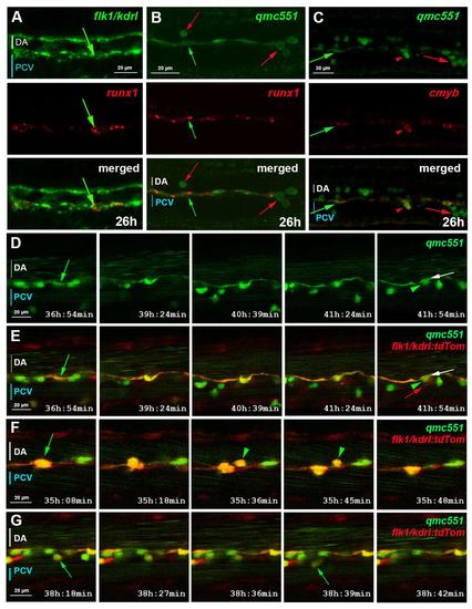
Fig. S2
- ID
- ZDB-FIG-160926-15
- Publication
- Thambyrajah et al., 2016 - A gene trap transposon eliminates haematopoietic expression of zebrafish Gfi1aa, but does not interfere with haematopoiesis
- Other Figures
- All Figure Page
- Back to All Figure Page
|
qmc551:GFP positive endothelial cells are haemogenic endothelial cells. This supplementary figure is related to Fig. 1. Confocal images of fixed (A-C) and live (D-G) embryos show optical sagittal sections through the DA with anterior left and dorsal up. The optical slices were at single cell resolution thickness, i.e. 2.0 (A,C), 1.0 (B) and 2.1 µm (D-G). Images in (D-G) are taken from timelapse experiments in which pictures were taken every 3 minutes. Times on panels represent hours and minutes after fertilization. (A) Double fluorescent runx1 and flk1/kdrl whole-mount in situ hybridisation (WISH). (B-C) Fluorescent runx1 (B) and cmyb (C) WISH combined with GFP immunohistochemistry. prRBCs (red arrows); prRBC progenitors in mesenchyme (red arrowheads) and HECs (green arrows). (D-G) Timelapse microscopy on qmc551;flk1:tdTom embryos from 31 hpf. (D) shows GFP expression only, while (E-G) present merged images. Annotations: HECs before (green arrow) and after bEMT (green arrowhead); DA endothelium after bEMT of HEC (white arrow); venous EC (red arrow). |
Reprinted from Developmental Biology, 417(1), Thambyrajah, R., Ucanok, D., Jalali, M., Hough, Y., Wilkinson, R.N., McMahon, K., Moore, C., Gering, M., A gene trap transposon eliminates haematopoietic expression of zebrafish Gfi1aa, but does not interfere with haematopoiesis, 25-39, Copyright (2016) with permission from Elsevier. Full text @ Dev. Biol.

