
Fig. 2
- ID
- ZDB-FIG-160714-1
- Publication
- Chang et al., 2016 - Leptospiral outer membrane protein LipL32 induces inflammation and kidney injury in zebrafish larvae
- Other Figures
- All Figure Page
- Back to All Figure Page
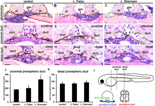
Leptospira Shermani induced acute tubular injury in zebrafish larvae. Transverse histological sections (H&E stain) from control (A,D,G), L. Patoc-treated (B,E,H), and L. Shermani-treated larvae (C,F,I). Note the marked swelling of the proximal pronephric ducts (circles) in L. Shermani-treated larvae compared with L. Patoc-treated larvae and controls. The differences were not evident in the distal pronephric ducts. Living larvae were incubated in E3 media containing L. Shermani or L. Patoc (1 × 10⁶ CFU/ml) from 24 hpf to 48 hpf. Control larvae were incubated with E3 buffer only. Representative micrographs from the level of the glomerulus (A-C), proximal pronephric duct (D-F), and distal pronephric duct (G-I) are shown. Circles indicate the location of the pronephric duct. Pt, pronephric tubule; Glm, glomerulus; Scale bar, 10 µm. (J,K) Quantification of the area of pronephric ducts. *P < 0.05 compared to control. n = 6 from three larvae in each group. (L) Diagram of transverse sections illustrating the structure of zebrafish pronephros at 48 hpf. |
| Fish: | |
|---|---|
| Condition: | |
| Observed In: | |
| Stage: | Long-pec |