
Fig. 3
- ID
- ZDB-FIG-160713-33
- Publication
- Chang et al., 2016 - Leptospiral outer membrane protein LipL32 induces inflammation and kidney injury in zebrafish larvae
- Other Figures
- All Figure Page
- Back to All Figure Page
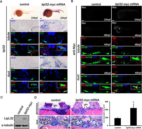
Microinjection of myc-tagged lipl32 mRNA and ectopic expression of LipL32 in zebrafish larvae. (A) Whole-mount in situ hybridization for lipl32 mRNA (lateral view, head to the left, scale bar, 200 µm). The expression of lipl32 mRNA was prominent in the head, the pronephric region (arrowhead) and the posterior blood island (arrow) in the transgenic wt1b:GFP line at 24 hpf. In transverse sections, signals detected by in situ hybridization are pseudocolored in red and merged to GFP immunostaining (green) to locate the pronephros. The white arrows indicate colocalization (orange stain) of lipl32 and GFP signals. Nuclei are stained with DAPI (blue). Scale bar, 20 µm. (B) Whole-mount immunostaining for Myc tag at 24 and 48 hpf. The expression of Myc tag was detected in the pronephric region (arrowhead) in lipl32 mRNA-injected larvae (scale bar, 200 µm). In transverse sections, the white arrow indicates colocalization (orange stain) of Myc tag (red) and wt1b:GFP fluorescence (green) in pronephric tubules and ducts. Scale bar, 10 µm. (C) Cropped western blot shows the expression of LipL32 protein in lipl32 mRNA-injected larvae at 48 hpf but not in control larvae under the same experimental condition. Whole larva lysates were immunoblotted with a customized antibody against LipL32. The uncropped blot is shown in Supplementary Figure S1. (D) Transverse sections (H&E stain, scale bar, 10 µm) show non-fusion of the glomerulus and markedly swelling of the pronephric ducts in lipl32 mRNA-injected larvae. Quantification of the area of proximal pronephric ducts is shown (*P < 0.05 compared to control). Pt, pronephric tubule; Glm, glomerulus. Circles indicate the location of the pronephric duct. Control larvae were injected with pCS2 (A,B) or myc mRNA (C,D). |