
Fig. 6
- ID
- ZDB-FIG-160714-3
- Publication
- Chang et al., 2016 - Leptospiral outer membrane protein LipL32 induces inflammation and kidney injury in zebrafish larvae
- Other Figures
- All Figure Page
- Back to All Figure Page
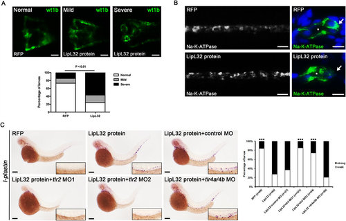
Injection of recombinant LipL32 protein induced pronephric kidney deformity, inflammation, and translocation of NA-K-ATPase. (A) The frequencies of deformities in pronephros were significantly increased in the LipL32-injected group compared to the control group (wt1b:GFP larvae at 48 hpf, n = 21 to 26 in each group, P < 0.01). Purified recombinant LipL32 protein was microinjected into the tail vein of zebrafish larvae at 24 hpf. Control larvae were injected with RFP. Photos were collected by in vivo observation under fluorescence microscopy (dorsal view, anterior to the left). Scale bar, 50 µm. (B) Immunostaining for NA-K-ATPase shows disruption of the basolateral location of NA-K-ATPase in the pronephric ducts in LipL32-injected larvae at 48 hpf. Right panels (scale bar, 5 µm) are the transverse sections of whole-mount stained larvae on the left (scale bar, 20 µm). Asterisks denote the lumens of pronephric ducts. Arrows indicate the basolateral cell surface. (C) In situ hybridization at 48 hpf shows that l-plastin-positive cells were increased after injection of LipL32 and the response was blocked by morpholino knockdown of tlr2 but not tl4a and tl4b. The insets are enlarged views of the corresponding regions of pronephric ducts. Scale bar, 200 µm. The diagram indicates the frequencies of strong and weak staining of l-plastin with different MOs (n = 27 to 101 from three experiments, ***P < 0.0001 versus LipL32 protein). |