
Fig. 1
- ID
- ZDB-FIG-160315-18
- Publication
- Govindan et al., 2016 - Cx43-Dependent Skeletal Phenotypes Are Mediated by Interactions between the Hapln1a-ECM and Sema3d during Fin Regeneration
- Other Figures
- All Figure Page
- Back to All Figure Page
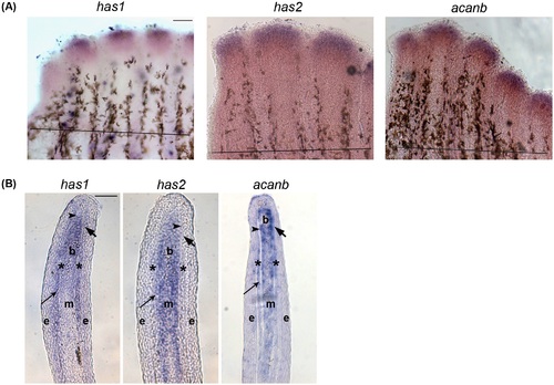
In situ hybridization showing the expression of Hapln1a-ECM components on 5 dpa regenerating fins. (A) Whole mount in situ hybridization of components of the Hapln1a-ECM. The genes has1, has2, and acanb are expressed during fin regeneration. The amputation plane is indicated by a black line in all the panels. Scale bar represents 100µm. (B) In situ hybridization on a WT 5 dpa cryo-section reveals compartmental expression of has1, has2, and acanb. Blastema (b), mesenchyme (m), and skeletal precursor cells (*). The thick arrow identifies the basal layer of the epidermis, which underlies the epidermis (e). The thin arrow identifies lepidotrichia and the arrowhead identifies the actinotrichia. Scale bar represents 50µm. |
| Genes: | |
|---|---|
| Fish: | |
| Condition: | |
| Anatomical Terms: | |
| Stage: | Adult |