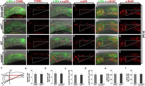
Chondrogenic cell survival and proliferation are unaffected in wls morphants at 24 hpf. (A–L) Confocal images showing lateral views of the heads of control-MO-injected (5mis-wlsMO) (A–F) or wls-MO-injected (wlsMO) (G–L) embryos stained by anti-Wls antibody (green in A,C,E,G,I,K) plus TUNEL (red in A,B,G,H), anti-pH3 (red in C,D,I,J) or anti-BrdU (red in E,F,K,L). The dotted lines of each triangle are defined as described in the Materials and Methods. The black circles indicate the position of the left eyes of the embryos. (M–X) Confocal images showing lateral view of the heads of WT (M–R) or wnt5b mutant (G–L) embryos stained by anti-Wls antibody (green in M,O,Q,S,U,W) plus TUNEL (red in M,N,S,T), anti-pH3 (red in O,P,U,V) or anti-BrdU (red in Q,R,W,X). (Y) The schematic on the left shows the three lines we defined to mark the region for counting the proliferating or apoptotic cells in the developing jaw (see Materials and Methods). (a–f) Statistical charts represent the quantitative results of apoptotic (TUNEL) or proliferating cells (pH3 or BrdU positive) at 24 hpf. There is no statistical difference between each set of these experiments. The n value is indicated. cMO, 5mis-wlsMO control.
|
