
Fig. S8
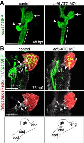
The effect of arf6 knockdown on the development of endoderm-derived organs. (A) Confocal images showing the endoderm and endoderm-derived organs at 48 hpf. The Tg(sox17:GFP) line was used to reveal the endoderm and endoderm-derived organs. Arrows, arrowheads, and open arrowhead point to the liver, the pancreas and the swim bladder, respectively; brackets mark the intestinal bulb. (B) Confocal images showing the hepatopancreatic ductal system at 75 hpf. Tg(Tp1:GFP);Tg(fabp10a:dsRed) larvae were processed for whole-mount immunostaining with 2F11 (gray), GFP (green), and dsRed (red) antibodies. In contrast to the intrahepatic biliary defect, the hepatopancreatic ductal system, revealed by 2F11 antibody, appeared to be normal in arf6-ATG MO-injected larvae. Arrows, arrowheads, open arrowheads, and open arrows point to the gallbladder (gb), the extrahepatic duct (ehd), the common bile duct (cbd), and the extrapancreatic duct (epd), respectively. The hepatopancreatic ductal system is schematically illustrated. Ventral views, anterior up. Scale bars, 50 µm. |
| Antibody: | |
|---|---|
| Fish: | |
| Knockdown Reagent: | |
| Anatomical Terms: | |
| Stage: | Protruding-mouth |
| Fish: | |
|---|---|
| Knockdown Reagent: | |
| Observed In: | |
| Stage Range: | Long-pec to Protruding-mouth |