
Fig. 10
- ID
- ZDB-FIG-150602-27
- Publication
- Liu et al., 2015 - Differential expression of protocadherin-19, protocadherin-17, and cadherin-6 in adult zebrafish brain
- Other Figures
- All Figure Page
- Back to All Figure Page
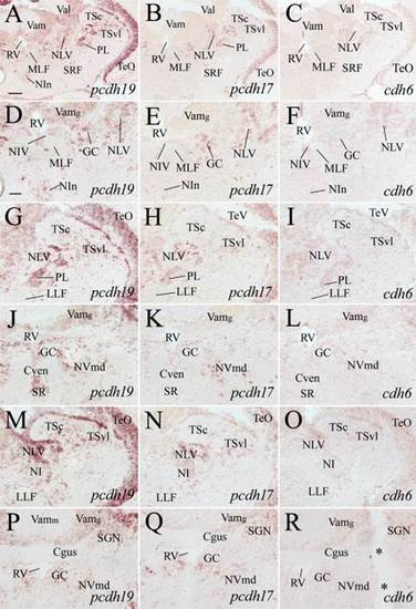
Expression of pcdh19, pcdh17, and cdh6 in the tegmentum, isthmus, cerebellar valvula, and anterior medulla. A-C: From adjacent sections from a level indicated by Fig. 1. D-F: Magnified views of the centromedial region, while G-I are higher magnifications of the dorsolateral region of their respective images in the top panels. J-L and M-O show similar respective regions from sections posterior (120-160 µm) to those shown in D-I. P-R: Higher magnifications of similar areas shown in J-L, but from sections 30-50 µm posterior to J-L. The two asterisks in R indicate a large artificial crack in the tissue. See list for abbreviations. Scale bar = 100 µm for A-C, 50 µm for the remaining panels (D-R have the same magnification). |