
Fig. 1
- ID
- ZDB-FIG-150319-5
- Publication
- Takeuchi et al., 2015 - Establishment of Gal4 transgenic zebrafish lines for analysis of development of cerebellar neural circuitry
- Other Figures
- All Figure Page
- Back to All Figure Page
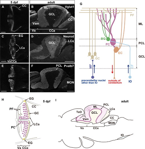
Cerebellar neural circuitry in zebrafish. (A–F) Expression of Vglut1 (A, B), Neurod (C, D), and parvalbumin7 (Pvalb7, E, F) in the cerebellar regions of larvae at 5 dpf (A, C, E, dorsal projection views, with anterior to the left) and adult sagittal sections (B, D, F, optical sections with anterior to the left). Vglut1 and Neurod signals mark the axons and somata of granule cells. Pvalb7 signals mark both neurites (axons and dendrites) and somata of Purkinje cells. (G) Schematic representation of cerebellar neural circuitry. (H, I) Schematic drawing of the cerebellum at 5 dpf (H, dorsal view) and of a sagittal section of the adult midbrain and hindbrain region. Scale bars: 50 µm in A, C, E; 200 µm in B, D, F. |
| Antibodies: | |
|---|---|
| Fish: | |
| Anatomical Terms: | |
| Stage Range: | Day 5 to Adult |
Reprinted from Developmental Biology, 397(1), Takeuchi, M., Matsuda, K., Yamaguchi, S., Asakawa, K., Miyasaka, N., Lal, P., Yoshihara, Y., Koga, A., Kawakami, K., Shimizu, T., Hibi, M., Establishment of Gal4 transgenic zebrafish lines for analysis of development of cerebellar neural circuitry, 1-17, Copyright (2015) with permission from Elsevier. Full text @ Dev. Biol.