
FIGURE
Fig. S4
- ID
- ZDB-FIG-150319-18
- Publication
- Takeuchi et al., 2015 - Establishment of Gal4 transgenic zebrafish lines for analysis of development of cerebellar neural circuitry
- Other Figures
- All Figure Page
- Back to All Figure Page
Fig. S4
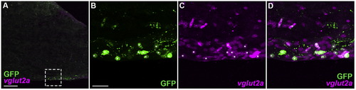
hspGFFDMC28C line marks glutamatergic neurons in the IO. Adult brain sagittal sections of hspGFFDMC28C; UAS:GFP were stained with vglut2a riboprobes (magenta) and anti-GFP antibody (green). (A) Low magnification view of the posterior hindbrain. (B, C, D) High magnification view of a box in A. Note that most of the GFP-positive cells are also vglut2a-positive (marked by asterisks). Scale bars: 100 µm in A; 20 µm in B (applied to B–D). |
Expression Data
Expression Detail
Antibody Labeling
Phenotype Data
Phenotype Detail
Acknowledgments
This image is the copyrighted work of the attributed author or publisher, and
ZFIN has permission only to display this image to its users.
Additional permissions should be obtained from the applicable author or publisher of the image.
Reprinted from Developmental Biology, 397(1), Takeuchi, M., Matsuda, K., Yamaguchi, S., Asakawa, K., Miyasaka, N., Lal, P., Yoshihara, Y., Koga, A., Kawakami, K., Shimizu, T., Hibi, M., Establishment of Gal4 transgenic zebrafish lines for analysis of development of cerebellar neural circuitry, 1-17, Copyright (2015) with permission from Elsevier. Full text @ Dev. Biol.