
Fig. 2
- ID
- ZDB-FIG-141222-23
- Publication
- Kizil et al., 2014 - Simplet/Fam53b is required for Wnt signal transduction by regulating β-catenin nuclear localization
- Other Figures
- All Figure Page
- Back to All Figure Page
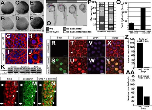
Smp regulates β-catenin nuclear localization. (A) Activity of the 7xTCF:mCherry transgene at 95% epiboly after mismatch morpholino injection, (B) knockdown of smp, (C) overexpression of β-catenin in mismatch controls and (D) overexpression of β-catenin in smp morphants. (E) Activity of the 7xTCF:mCherry transgene at 95% epiboly after overexpression of lef1-VP16 in mismatch controls and (F) overexpression of lef1-VP16 in smp morphants. (G,H) Immunohistochemistry staining for β-catenin (red) and staining of nuclei with DAPI (blue) in mismatch control and (I,J) smp morphants. (K) Western blots for β-catenin in nuclear and cytoplasmic lysates from mismatch-morpholino controls (mm-mo) and smp morphants (as-mo). Total amount of β-catenin levels in the cells was unaltered by the smp knockdown. Loading controls were γ-tubulin for the cytoplasmic fraction and H2A for the nuclear fraction. (L) Immunohistochemistry staining for zebrafish Smp shows positive nuclei in the marginal zone where Wnt signaling is active. (M) Immunohistochemistry staining for β-catenin shows localization at the plasma membrane and in distinct nuclei (white arrowheads). (N) Merged stainings show colocalization of Smp and β-catenin in nuclei (white arrowheads) and cells with Smp in nuclei lacking β-catenin (white arrows). (O) Overexpression of wnt8 during zebrafish development produces phenotype classes that affect normal development of eyes (blue arrow); the midbrain-hindbrain boundary (pink arrow); and the somites and posterior structures (green arrows). (P) Percentage occurrence of phenotypic classes produced by overexpression of wnt8 alone or overexpression of wnt8 with smp. (Q) Results of luciferase assays of either the pBAR reporter (β-catenin binding sites) or the pFuBAR reporter (mutated β-catenin sites) for Wnt activity in HEK293T cells. (R) Uninjected controls. (S) GFP localization in the nuclei of embryos injected with mRNA encoding Smp-GFP. (T) β-Catenin localization in the dorsal region of control embryos. (U) β-Catenin localization in Smp-GFP-injected embryos. (V,W) DAPI staining labels nuclei. (X,Y) Merged fluorescence for β-catenin, GFP and DAPI. (Z) The number of β-catenin-positive cells with Smp in the nucleus or in the cytoplasm. (AA) The subcellular distribution of Smp in cells lacking β-catenin nuclear staining. Scale bars: 10μm in A-G,I,L-N; 1μm in H,J; 300μm in O; 10μm in R-Y. All experiments were performed at least three times. Data represent the mean; error bars indicate s.d. |
| Gene: | |
|---|---|
| Fish: | |
| Knockdown Reagents: | |
| Anatomical Term: | |
| Stage: | 90%-epiboly |