
Fig. S6
- ID
- ZDB-FIG-150116-3
- Publication
- Kizil et al., 2014 - Simplet/Fam53b is required for Wnt signal transduction by regulating β-catenin nuclear localization
- Other Figures
- All Figure Page
- Back to All Figure Page
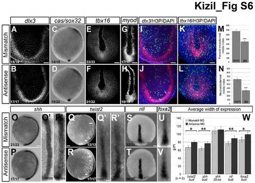
Gastrulation markers at tailbud stage are unaffected by loss of smp, Related to Figure 1. (A) Mismatch-morpholino-control embryos show dlx3b expression in the neuroectoderm. (B) dlx3b expression is similar in smp morphants (Antisense). (C) cassanova/sox32 (cas/sox32) is observed in developing endoderm in mismatch controls. (D) smp morphants display similar (cas/sox32) expression. (E) tbx16 expression in the axial and paraxial mesoderm of control embryos. (F) smp morphants display similar tbx16 expression. (G) myod staining in control embryos shows expression in the adaxial of the paraxial mesoderm. (H) smp morphants show similar myod expression. (I) Double staining for H3P and dlx3b. (J) Double staining for H3P and dlx3b in smp morphants show decrease in the number of H3P and dlx3b expression. (K) Double staining for H3P and tbx16 expression in controls. (L) Double staining for H3P and tbx16 in smp morphants. (M) Graph of the number of H3P-positive cells overlapping with confocal planes containing dlx3b expression. (N) Graph showing number of H3P-positive cells that overlap with confocal planes showing tbx16 expression. (O) Lateral view of shh expression in the developing prechordal plate of controls. (O′) Dorsal view of shh expression in control embryos. (P) Lateral view of shh expression in smp morphants show expression along the length of the AP axis. (P′) Dorsal view of shh expression in smp morphants shows that that its expression is wider than shh expression controls. (Q) Lateral view of twist2 expression in the developing prechordal mesoderm in controls. (Q′) Dorsal view of twist2 expression in prechordal plate of control embryos. (R) twist2 expression in smp morphants. (R′) Dorsal view of twist2 in smp morphants. (S) Dorsal view of early ntl expression in the developing prechordal plate. (T) Dorsal view of ntl expression in smp morphants. (U) Dorsal view of expression of foxa2 in prechordal plate of controls. (V) Dorsal view of foxa2 expression in smp morphants. (W) Measurements of the lateral dimensions of the prechordal plate genes show slightly increased dimensions of the prechordal plate in smp morphants and that this alteration is temporary based on measurements of the dimensions of shh expression at 5 somite embryos. Numbers in the lower left corners in panels indicate the number of embryos with the depicted expression patterns to the total number of embryos. Scale bars: 50 μm (A,B,E,F,I-L), 100 μm (C,D,O-T), 250 μm (G, H, O′-R′,U′,V′). |