Fig. 3
- ID
- ZDB-FIG-141125-4
- Publication
- Choksi et al., 2014 - Systematic discovery of novel ciliary genes through functional genomics in the zebrafish
- Other Figures
- All Figure Page
- Back to All Figure Page
|
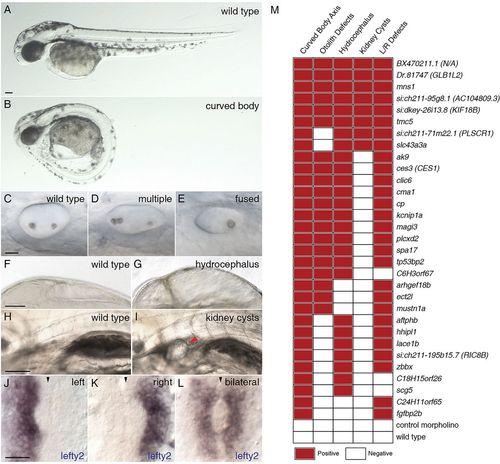
Morpholino knockdown of FIGs causes multiple ciliary dysfunction-associated phenotypes in zebrafish embryos. Five ciliary phenotypes were scored in the zebrafish embryos injected with morpholinos targeting the 50 selected FIGs. The extent of body axis curvature: (A) wild type or (B) curved body. Otolith defects in the inner ear: (C) two otoliths, (D) greater than two otholiths or (E) a single otolith. Swelling of the brain ventricles (hydrocephalus): (F) normal or (G) hydrocephalus. Kidney cysts: (H) no cysts or (I) a minimum of one cyst per embryo. Expression of lefty2: (J) leftward expression, (K) rightward expression or (L) bilateral expression or no expression (data not shown). The red arrowhead indicates the position of a kidney cyst (I); the black arrowheads indicate the midline (J-L). Scale bars: 100μm in A,F,H; 20μm in C,J. (M) Morpholino knockdowns of 31 genes exhibited defects in at least two of the assayed phenotypes. For morphological phenotypes, each square represents at least 60 assayed embryos from two independent injections. For left-right asymmetry defects, each square represents a minimum of 30 stained and scored embryos. A red square indicates that the morphant phenotype level was significantly different from the wild type and met a minimum defective percentage (see Materials and Methods). |

