
Fig. 6
- ID
- ZDB-FIG-141106-3
- Publication
- Connors et al., 1999 - The role of tolloid/mini fin in dorsoventral pattern formation of the zebrafish embryo
- Other Figures
- All Figure Page
- Back to All Figure Page
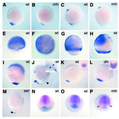
tld expression pattern and altered tld, chd and bmp4 expression in mutant embryos. eve1 expression in bud stage wild-type (A) and mfn mutant (B) embryos. bmp4 expression in 1-somite-stage wild-type (C) and mfn mutant (D) embryos. tld expression in wild-type embryos at 55% epiboly (E), 70% epiboly (F,G), 85% epiboly (H), bud-stage (I) and 20-somite-stage (J) wild-type embryos. Arrowhead in J points to tld expression in the presumptive vasculature. Inset in J is a higher magnification showing the posterior trunk of a slightly older embryo with tld expression marking the prospective artery and vein (arrows). tld expression at 5 somites in wild-type (K), chordino (L) and swirl (M) mutant embryos. Insets in K-L are higher magnification caudal views of tld expression, anterior to the top. Reduced tld expression (arrowhead) in a swirl mutant. Double in situ of chd (magenta) and bmp4 (blue) expression in a bud-stage wild-type embryo (N). Double in situ of chd (magenta) and tld (blue) expression in bud-stage wild-type (O) and mfn mutant (P) embryos. (P) Expanded chd expression (arrows) in a mfn mutant. (A-D, G-M) Lateral views, dorsal to the right. (E) Dorsal view, slightly tilted downward. (F) Animal pole view. (N-P) Vegetal pole view, anterior/dorsal to the top. |
| Genes: | |
|---|---|
| Fish: | |
| Anatomical Terms: | |
| Stage Range: | 50%-epiboly to 20-25 somites |