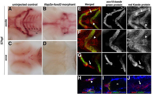
Zebrafish craniofacial tendon populations are derived from the neural crest. Morpholino-mediated knockdown of tfap2a and foxd3 results in (A,B) complete loss of sox9a-positive pharyngeal cartilage (98%, n=58) and (C,D) scxa-positive craniofacial tendon progenitors (96%, n=46) at 57hpf compared with controls. (E-G) 72hpf photoconverted sox10:kaede embryos express sox10:kaede green protein in the pharyngeal cartilage (E-G and middle panel), and the red Kaede protein from the 22hpf photoconversion is found in the two major populations of craniofacial tendon progenitors (E, arrow and arrowhead; F,G, right panel). (H-J) Colocalization of scxa and Kaede protein in photoconverted sox10:kaede embryos at 72hpf is observed in the sternohyoideus connection point (H,I; sternohyoideus muscle is labeled m) and in the ligament (J). A subset of the scxa-positive and Kaede-positive cells also co-expresses xirp2a transcripts. Arrows in E,G,J mark ligament medial to palatoquadrate; arrowheads in E,F,H mark tendon connecting the sternohyoideus to the ceratohyals. cb: ceratobranchials; ch, ceratohyal; hs, hyosymplectic; m, muscle; mc, Meckel′s cartilage; ne, neurocranium; pq, palatoquadrate.
|
