
FIGURE
Fig. S2
- ID
- ZDB-FIG-140515-8
- Publication
- Weber et al., 2014 - Mitotic Position and Morphology of Committed Precursor Cells in the Zebrafish Retina
- Other Figures
- All Figure Page
- Back to All Figure Page
Fig. S2
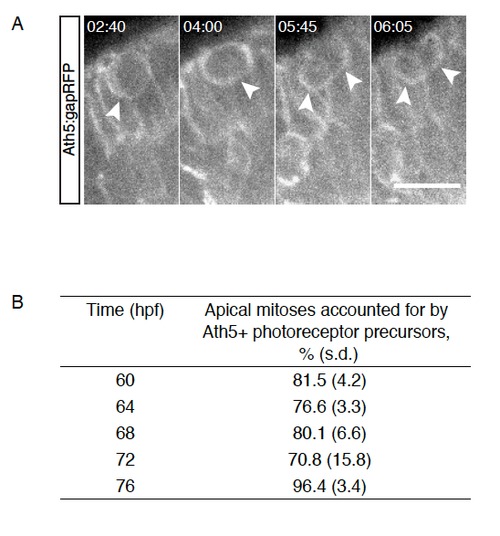
Late apical divisions give rise to photoreceptors, Related to Figure 3 A) Tg(Ath5:gapRFP); An Ath5+ cell divides within the PR layer and gives rise to two Ath5+ daugther cells. Time is hr:min. Imaging started at 60hpf. B) Percentage of apical mitoses accounted for by Ath5+ cells dividing within the PR layer between 60hpf and 76hpf (4h intervals). n=2-3 embryos/ time point. |
Expression Data
Expression Detail
Antibody Labeling
Phenotype Data
Phenotype Detail
Acknowledgments
This image is the copyrighted work of the attributed author or publisher, and
ZFIN has permission only to display this image to its users.
Additional permissions should be obtained from the applicable author or publisher of the image.
Full text @ Cell Rep.