
Fig. 2
- ID
- ZDB-FIG-140515-3
- Publication
- Weber et al., 2014 - Mitotic Position and Morphology of Committed Precursor Cells in the Zebrafish Retina
- Other Figures
- All Figure Page
- Back to All Figure Page
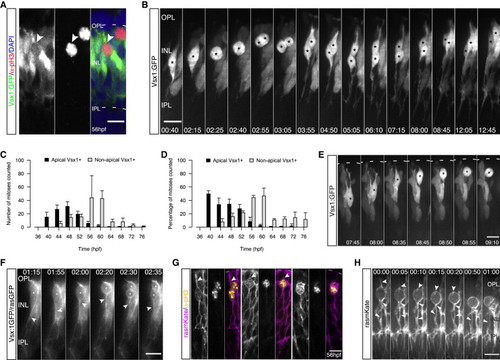
Nonapically Dividing Monopolar Vsx1+ Precursors Give Rise to Two Bipolar Cells (A) pH3 (red) and DAPI staining (blue) in a Vsx1:GFP (green) embryo. pH3+/Vsx1+ cells are observed in the INL (arrows). (B) Two Vsx1+ cells arise from a nonapically dividing Vsx1:GFP cell, align in the INL, and feature BC morphology (asterisk) (from Movie S2). Time hr:min. Imaging started 50 hpf. (C) Time course showing counts of apically and nonapically dividing Vsx1+ cells/embryo (36–76 hpf; 4 hr intervals). Error bars represent SD; n = 2–3 embryos per time point. (D) Data from (C) as percentage of all counted mitoses. Error bars represent SD. (E) Vsx1:GFP cell bodies undergo IKNM-like movement prior to subapical division (asterisk). Time in hr:min. Imaging started at 48 hpf. (F) A transplanted Vsx1:GFP/rasGFP cell divides adjacent to the OPL (asterisk), featuring a basal process before, during, and after mitosis (notched arrows). Time in hr:min. Imaging started at 52 hpf. (G) Embryos mosaically injected with rasmKate RNA (magenta) stained for pH3 (yellow). Cells are attached apically in late G2 (left, arrows). An apical process is present at the beginning of cell rounding (arrows, middle) but lost upon entry into mitosis (right). (H) Time-lapse of an embryo mosaically injected with rasmKate. Upon onset of cell rounding, an apical process can be observed (notched arrows), which is lost upon entry into mitosis. Filled arrows point toward basal process; from Movie S3. Time in hr:min. Imaging started at 56 hpf. Scale bars represent 10 μm. See also Figure S1. |