
Fig. S1
- ID
- ZDB-FIG-140515-7
- Publication
- Weber et al., 2014 - Mitotic Position and Morphology of Committed Precursor Cells in the Zebrafish Retina
- Other Figures
- All Figure Page
- Back to All Figure Page
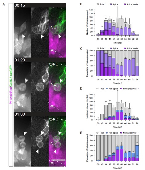
A) HC precursors dividing in the INL are non-polar, Related to Figure 1. Live imaging of Ptf1A:dsRed/Cx55.5:rasGFP double transgenic embryos. Time in hr:min. Imaging started at 48hpf. B)-E) Time-courses showing the absolute and relative number and location of Vsx1+ divisions, Related to Figure 2. B) Counts of apical Vsx1+ divisions (magenta) overlaid on counts of all apical divisions (light purple) and counts of all divisions (grey). C) Apical Vsx1+ divisions (magenta) and apical divisions (light purple) as a percentage of all divisions counted. D) Counts of non-apical Vsx1+ divisions (magenta) overlaid on counts of all non-apical divisions (light blue) and counts of all divisions (grey). E) Non-apical Vsx1+ divisions (magenta) and non-apical divisions (light purple) as a percentage of all divisions counted. For all graphs shown n=2-3 embryos/time point and error bars = SD. |