
Fig. 2
- ID
- ZDB-FIG-140422-40
- Publication
- Holly et al., 2014 - Sfrp1a and Sfrp5 function as positive regulators of Wnt and BMP signaling during early retinal development
- Other Figures
- All Figure Page
- Back to All Figure Page
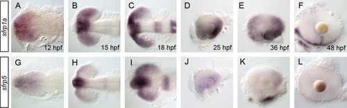
sfrp1a and sfrp5 mRNAs are expressed in the eye during retinal development. The spatio-temporal pattern of secreted frizzled related protein (sfrp) mRNA expression was assessed using whole mount in situ during zebrafish eye development. Initial expression of sfrp1a demarcates the presumptive eye field (A,B) with later stages displaying patterns specific to the presumptive ventral retina (C–E). Expression of sfrp1a persists in the ventral retina up to 48 hpf where it becomes constrained to the choroid fissure (F). In contrast, sfrp5 expression is more ventrally restricted early (G–I), then decreases significantly from 25 hpf and is absent from the retina by 48 hpf (J–L). Embryos shown are flat-mounted in dorsal (A–C,G–I) or lateral views of dissected eyes (D–F, J–L). |
| Genes: | |
|---|---|
| Fish: | |
| Anatomical Terms: | |
| Stage Range: | 5-9 somites to Long-pec |
Reprinted from Developmental Biology, 388(2), Holly, V.L., Widen, S.A., Famulski, J.K., and Waskiewicz, A.J., Sfrp1a and Sfrp5 function as positive regulators of Wnt and BMP signaling during early retinal development, 192-204, Copyright (2014) with permission from Elsevier. Full text @ Dev. Biol.