
Fig. 3
- ID
- ZDB-FIG-140422-41
- Publication
- Holly et al., 2014 - Sfrp1a and Sfrp5 function as positive regulators of Wnt and BMP signaling during early retinal development
- Other Figures
- All Figure Page
- Back to All Figure Page
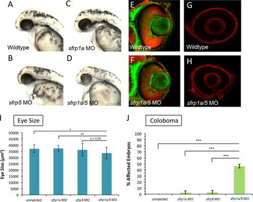
sfrp1a and sfrp5 depletion leads to small eyes and coloboma. To ascertain the function of Sfrp proteins during retinal development, we injected one-cell stage zebrafish embryos with morpholinos (MO) targeting sfrp1a and/or sfrp5. Embryos injected singly with 3 ng of sfrp1a or sfrp5 MO display overtly normal eye size at 25 hpf (A–C). In contrast, sfrp1a/5 MO co-injected embryos (3 ng each MO) display smaller eyes (D, I) (N, p<0.05; NN, p<0.01, ANOVA). 48 hpf retinas stained with phalloidin and TO-PRO3 display no overt difference between wildtype and sfrp1a/5 MO co-injected embryos (E–F). At 48 hpf WT embryos display a dissolution of Laminin staining at the choroid fissure (G). sfrp1a/5 MO embryos display persistent Laminin staining at the choroid fissure, indicating a coloboma phenotype (G,H). The increase in observed coloboma frequency is statistically significant (NNN, p<0.0036, Fisher′s Exact) when compared with wild type or embryos injected singly with Sfrp MOs (J). |
| Antibody: | |
|---|---|
| Fish: | |
| Knockdown Reagents: | |
| Anatomical Term: | |
| Stage: | Long-pec |
| Fish: | |
|---|---|
| Knockdown Reagents: | |
| Observed In: | |
| Stage Range: | Prim-5 to Long-pec |
Reprinted from Developmental Biology, 388(2), Holly, V.L., Widen, S.A., Famulski, J.K., and Waskiewicz, A.J., Sfrp1a and Sfrp5 function as positive regulators of Wnt and BMP signaling during early retinal development, 192-204, Copyright (2014) with permission from Elsevier. Full text @ Dev. Biol.