|
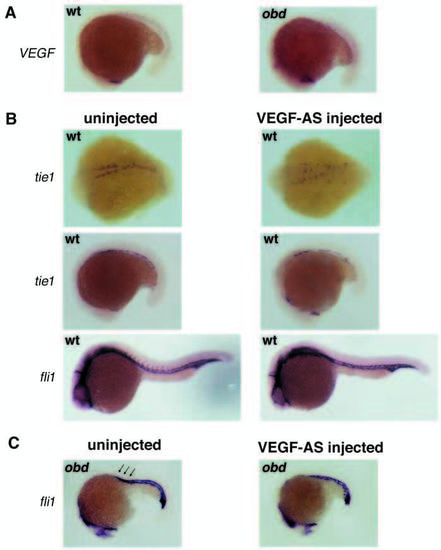
VEGF antisense delays precocious migration of obd angioblasts. (A) VEGF is normally expressed segmentally, beginning at the 17 somite stage, in both wild-type and obd embryos. (B) In a wild-type untreated embryo at 14 somites, angioblasts have migrated from the LPM to the midline while in a 1 mM VEGF-antisense treated embryo, tie1-positive cells are still localized laterally (top row). The dorsal aorta and vein are formed in both treated and untreated embryos at 19 somites, although the vessels in the injected embryo are discontinuous (middle row). At 24 hours, angiogenic sprouts are evident in an untreated embryo but are not seen in the treated embryos (bottom row). (C) The ectopic sprouts evident in a 19 somite obd embryo (arrows), are completely inhibited in the VEGF-AStreated obd mutant embryo.
|

