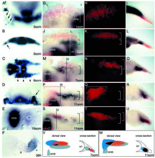
Cardiac expression of fgf8 in relation to heart marker genes in wild-type embryos. (A-F) fgf8 expression at various stages of development, as indicated. B and D are cross sections of A and C, respectively. (A,B) Cells in lateral plate mesoderm express fgf8 (arrows) at the 3-somite stage. (C,D) Expression bilaterally to the neural tube including cardiogenic fields (arrowheads in C and arrow in D) at the 8-somite stage. (E) Ring-shaped expression in the heart at 19-somite stage. (F) Expression is predominantly in the ventricle at 36 hpf. (F) Dissected heart. (A) Anterior to the top; (C,E) anterior to the left; a, atrium; h, heart; mhb, mid-hindbrain boundary; os, optic stalks; r2, rhombomere 2; r4, rhombomere 4; v, ventricle. (G-U) Double in situ hybridization with fgf8 (black) and indicated heart markers (red fluorescence) of wild-type embryo at given stages. (G-I) fgf8 is expressed in close proximity to nkx2.7. (J-L) fgf8 is expressed close to gata4. (M-O) fgf8 expression partially overlaps nkx2.5 expression (star marks the same cell in M and N). (P-R) fgf8 expression partially overlaps gata4 expression. (S-U) fgf8 expression strongly overlaps nkx2.5 expression. Brackets indicate the two expression domains. (V,W) Summary of fgf8 expression relative to the described heart marker genes at given stages (grey, fgf8; red, nkx2.5; blue, gata4, black, triple overlap). Embryos in G,H,J,K,M,N,P,Q,S,T are flat mounted, anterior to the left; G,J,M,P,S are bright-field images, H,K,N,Q,T are fluorescent images of the same embryos; I,L,O,R,U are cross sections at the indicated levels, lateral is to the right. mhb, mid-hindbrain boundary; r4, rhombomere 4.
|
