Fig. 9
- ID
- ZDB-FIG-140304-44
- Publication
- Gariano et al., 2014 - Analysis of three mu-AP1 subunits during zebrafish development
- Other Figures
- All Figure Page
- Back to All Figure Page
|
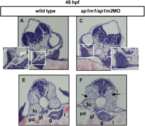
A–F: Histological analysis of double morphants. At 48 hpf zebrafish ap1m1/ap1m2MO double knockdown embryos were embedded in paraffin and sectioned at trunk level (H&E staining). Transverse sections (7μm) revealed disturbed pronephritic duct development (C,D; controls A,B); (see boxed areas in panel A and C). Structures in the anterior part as pronephric ducts, gut, liver in control (E) and double morphants (F). In double morphants the lumen of gut and the cell polarity are lost, liver morphology is severely compromised, the pronephric ducts are not well formed (F). An altered myotome organization is also detected (F, black arrows; control E). Abbreviations: pd, pronephric duct; g, gut; tu, tubule; gl, glomerule; l, liver. |
| Fish: | |
|---|---|
| Knockdown Reagents: | |
| Observed In: | |
| Stage: | Long-pec |

