
Fig. 5
- ID
- ZDB-FIG-140304-40
- Publication
- Gariano et al., 2014 - Analysis of three mu-AP1 subunits during zebrafish development
- Other Figures
- All Figure Page
- Back to All Figure Page
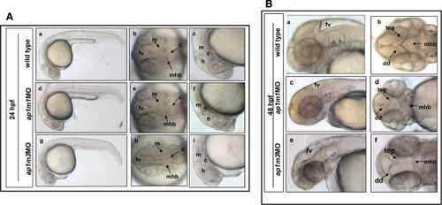
Morphological defects in ap1m1MO and ap1m3MO single morphants. Embryos were injected at the two-cell stage and examined at 24 and 48 hpf. A: Lateral view and dorsal view at 24 hpf. Single morphants of ap1m1 and ap1m3 exhibit brain malformations (d,e,g,h) with unclear boundaries between brain subdivisions, especially at hindbrain and midbrain region (f,i). ap1m3MO morphants showed poorly defined somites (g). B: At 48 hpf, CNS abnormalities became more pronounced. The forebrain ventricle in both single morphants (c,e; control in a). Dorsal view of ap1m1MO and ap1m3MO embryos revealed a severe disorganization of CNS structures (d,f). a,c,e: lateral view anterior to the left. b,d,f: dorsal view. h, hindbrain; c, cerebellum; teg, tegmentum; dd, dorsal diencephalon; mhb, midbrain–hindbrain boundary; m, midbrain; fv, forebrain ventricle. |
| Fish: | |
|---|---|
| Knockdown Reagents: | |
| Observed In: | |
| Stage Range: | Prim-5 to Long-pec |