Fig. 4
- ID
- ZDB-FIG-140304-39
- Publication
- Gariano et al., 2014 - Analysis of three mu-AP1 subunits during zebrafish development
- Other Figures
- All Figure Page
- Back to All Figure Page
|
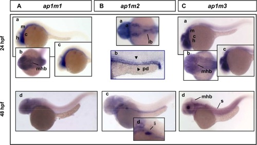
Expression analysis of zebrafish μ1 adaptins by WISH at 24 and 48 hpf. A: ap1m1 is expressed in defined brain regions at 24 hpf (a,b c) and 48 hpf (d). a,c,d: lateral view anterior to the left. b: dorsal view B: (a–d) ap1m2 transcript is expressed in intestinal bulb (a) and pronhephric ducts at 24 hpf (b). At 48 hpf the transcript expression is still detectable in the digestive tract (c) and liver (d). b,c,d lateral view anterior to the left; a: dorsal view. C: (a–d) The ap1m3 transcript is detectable at 24 (a,b,c) and 48 hpf (d). Expression in different CNS structures: midbrain, hindbrain and cerebellum (a) and midbrain–hindbrain boundary (d). At 48 hpf a label of ap1m3 is detectable in somites (d). m, midbrain; h, hindbrain; mhb, midbrain–hindbrain boundary; c, cerebellum; s, somites; pd, pronephric duct; ib, intestinal bulb; li, liver. |
| Genes: | |
|---|---|
| Fish: | |
| Anatomical Terms: | |
| Stage Range: | Prim-5 to Long-pec |

