
Fig. 2
- ID
- ZDB-FIG-140122-23
- Publication
- Venkiteswaran et al., 2013 - Generation and Dynamics of an Endogenous, Self-Generated Signaling Gradient across a Migrating Tissue
- Other Figures
- All Figure Page
- Back to All Figure Page
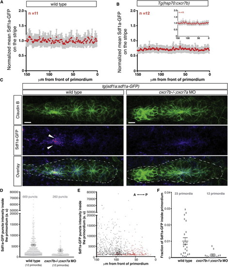
Cxcr7 Is Required for Sdf1a-GFP Sequestration by the Primordium (A and B) Average fluorescence intensity of Sdf1a-GFP protein along the stripe of chemokine-producing cells underneath primordia in embryos of indicated genotypes (B, inset, heat-shocked wild-type control embryos). (C) Sum projection of the primordium from 36 hpf embryos of indicated genotypes stained for Claudin-B and GFP. Scale bar, 10 μm. Direction of migration is to the right. Arrowheads indicate Sdf1a-GFP puncta. (D) Distribution of intensities of Sdf1a-GFP puncta in primordia of indicated genotypes. (E) Distribution of intensities of Sdf1a-GFP puncta in primordia along the anterior-posterior axis. Each dot (D and E) represents an individual punctum (red, cxcr7b/; cxcr7a morphant primordia; black, wild-type primordia). (F) Fraction of Sdf1a-GFP found inside of the primordium of the total Sdf1a-GFP in the indicated genotype. Each dot represents the fraction of Sdf1a-GFP in an individual primordium. Horizontal bars represent the mean ± SEM. See also Figure S1 and Data S1 and S2. |
| Gene: | |
|---|---|
| Fish: | |
| Knockdown Reagents: | |
| Anatomical Terms: | |
| Stage: | Prim-25 |
Reprinted from Cell, 155(3), Venkiteswaran, G., Lewellis, S.W., Wang, J., Reynolds, E., Nicholson, C., and Knaut, H., Generation and Dynamics of an Endogenous, Self-Generated Signaling Gradient across a Migrating Tissue, 674-687, Copyright (2013) with permission from Elsevier. Full text @ Cell