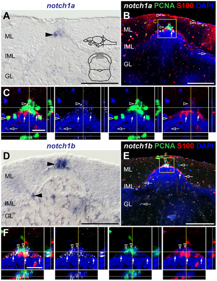
notch1a and notch1b expression in the adult cerebellar niche.
Cross-sections at the indicated level through the mesencephalon; cerebellar area shown in the micrographs is indicated in the cross-section schematic in A. Brightfield images show expression of A, notch1a and D, notch1b in the cerebellum (arrowheads). Confocal images showing localization of the glial marker S100β (red) and PCNA (green), with B-C, notch1a and E-F, notch1b by FISH (white). B-C, Expression of notch1a localizes with a large fraction of PCNA+ cells in the niche (unfilled arrowheads); weak (white arrow) or no expression of notch1a is detected in the Bergmann glia (S100β+ ). E-F, notch1b is expressed in a subpopulation of PCNA cells (unfilled arrowheads) and in a few S100β+ cells (white arrows). A few scattered notch1a+ and notch1b + cells in the ML, IML and GL do not localize with the analysed markers (unfilled arrows). Abbreviations: GL, granule cell layer; IML, intermediate layer; ML, molecular layer. Scale bars = 50 μm in A, B, D and E; 20 μm in C and F.
|
