
Fig. S5
- ID
- ZDB-FIG-140114-6
- Publication
- de Oliveira-Carlos et al., 2013 - Notch receptor expression in neurogenic regions of the adult zebrafish brain
- Other Figures
- All Figure Page
- Back to All Figure Page
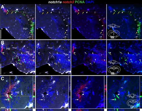
Overlapping and complementary notch1a/3 expression in the adult zebrafish hypothalamus. Confocal images of double FISH showing the localization of notch1a (white), notch3 (red), and PCNA (green). Cross-sections at the indicated level through the diencephalon; hypothalamic area shown in the micrographs is indicated in the cross-section schematics. A–C, notch1a is expressed in a subpopulation of notch3 + /PCNA + cells in Hv, Hd and Hc (filled white arrowheads); yellow arrows indicate Notch receptor - /PCNA + cells; unfilled yellow arrowheads indicate cells expressing notch3 alone. notch1a expression partially overlaps with the notch3 + /PCNA - population (unfilled white arrowheads); there are a few notch1a + /notch3 - /PCNA cells in Hv and Hd (filled white arrows). Abbreviations: Hc, caudal zone of the periventricular hypothalamus; Hd, dorsal zone of the periventricular hypothalamus; Hv, ventral zone of the periventricular hypothalamus. Scale bar = 100 μ in A (applies to B). |