|
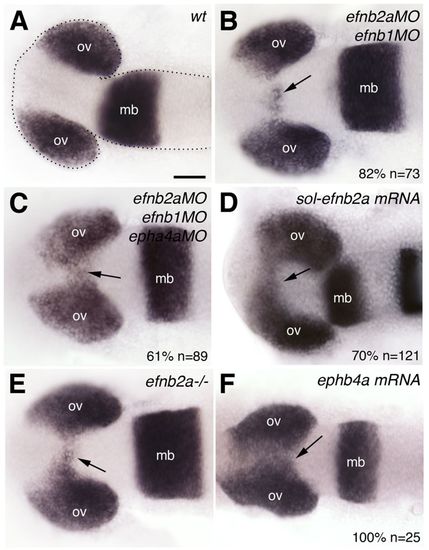
Partial abrogation of Eph/Ephrin activity interferes with optic vesicle evagination. Whole-mount in situ hybridisation in 10-12 ss wild-type zebrafish embryo (A), embryos injected with Eph/Ephrin morpholinos (B,C) or mRNA (D,F), and efnb2a mutant (E). The optic vesicles are labelled by expression of mab21/2. Arrows (B-F) indicate the presence of eye fated cells (labelled by mab21/2) embedded within the forebrain. All panels show dorsal views with anterior to the left. The phenotype shown in C is representative of 61% of the embryos analysed; the remaining 39% are not wild type but show a milder phenotype than that illustrated. The phenotype shown in F is representative for EphA4a and EphB4a misexpressions (epha4a: 70%, n=56; ephb4a: 100%, n=25). mb, midbrain; ov, optic vesicle. Scale bars: 50 μm.
|

