|
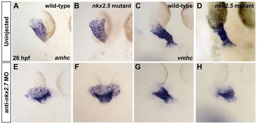
Loss of nkx2.7 function in nkx2.5 mutants exacerbates defects during heart tube extension. (A-H) In situ hybridization depicts expression of amhc (A,B,E,F) and vmhc (C,D,G,H) in wild-type embryos (A,C), nkx2.5 mutants (B,D), wild-type embryos injected with anti-nkx2.7 MO (E,G) and Nkx-deficient embryos (F,H). Dorsal views, anterior to the top, at 26 hpf. The nkx2.5 mutant embryos exhibit subtle defects in heart tube extension, including a broader atrial region (A,B) and a slightly shorter ventricular region (C,D). Following MO injection, wild-type embryos demonstrate a spread of atrial cells and a compact coalescence of the ventricular cells (E,G). MO injection into nkx2.5 mutants leads to an exacerbated phenotype with a sprawling, widened atrial portion and a stunted ventricular portion of the heart tube (F,H).
|