
Fig. 8
- ID
- ZDB-FIG-130906-30
- Publication
- Sorrell et al., 2013 - Tcf7l1 proteins cell autonomously restrict cardiomyocyte and promote endothelial specification in zebrafish
- Other Figures
- All Figure Page
- Back to All Figure Page
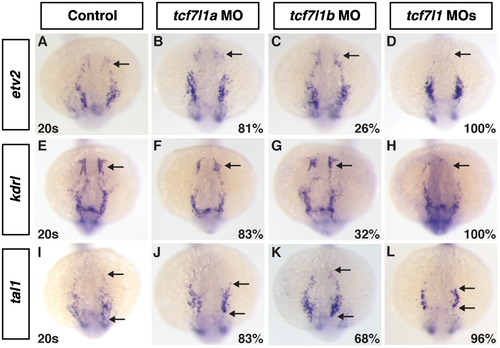
Anterior vasculature is absent in Tcf7l1 deficient embryos. ((A)–(D)) Etv2, ((E)–(H)), kdrl ((I)–(J)) tal1 expression at the 20s stage in control, Tcf7l1a, Tcf7l1b and Tcf7l1 deficient embryos. In Tcf7l1a and Tcf7l1b depleted embryos, there is a reduction in the anterior ECs ((B), (C), (F), (G)), with the defects in Tcf7l1a deficient embryos being more penetrant and stronger. Tcf7l1 deficient embryos have a complete loss of the ACEs ((D) and (H)). The more posterior expression of tal1 is reduced in Tcf7l1 deficient embryos compared to the individually depleted embryos. Images are dorsal with anterior up. Arrows in (A)–(H) indicate the ACEs. Arrow in (I)–(L) indicates the anterior and posterior limits of the tal1 expressing cells. |
| Genes: | |
|---|---|
| Fish: | |
| Knockdown Reagents: | |
| Anatomical Term: | |
| Stage: | 20-25 somites |
Reprinted from Developmental Biology, 380(2), Sorrell, M.R., Dohn, T.E., D'Aniello, E., and Waxman, J.S., Tcf7l1 proteins cell autonomously restrict cardiomyocyte and promote endothelial specification in zebrafish, 199-210, Copyright (2013) with permission from Elsevier. Full text @ Dev. Biol.