
Fig. 2
- ID
- ZDB-FIG-130821-6
- Publication
- Anderson et al., 2013 - Hepatocyte Growth Factor Signaling in Intrapancreatic Ductal Cells Drives Pancreatic Morphogenesis
- Other Figures
- All Figure Page
- Back to All Figure Page
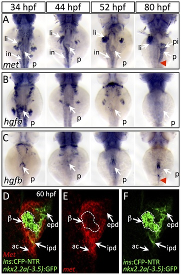
Dynamic expression of met and hgf during pancreas morphogenesis. (A) met is expressed strongly in the pancreatic bud (p), liver bud (li), and intestinal bulb (in) from 34–52 hpf. At 80 hpf, met expression is found throughout the exocrine pancreas and is diminished in the principal islet (pi). (B,C) hgfa (B) and hgfb (C) are expressed at 34–44 hpf near the dorsal pancreatic bud prior to pancreatic tail formation. At 52 hpf, at the onset of pancreatic tail formation, both hgfa and hgfb are expressed near the distal tip of the pancreatic bud, with hgfb expression persisting until the completion of pancreatic tail outgrowth (red arrowhead). (D–F) One-micron confocal plane images of duct:GFP;ins:CFP-NTR embryos stained for met mRNA using fluorescent in situ hybridization shows met expression (red) in both acinar cells (ac) and intrapancreatic ducts (ipd; green), but not in the principal islet (green outlined in white). Dorsal views, anterior to the top. beta-cells, β epd, extrapancreatic duct. |
| Genes: | |
|---|---|
| Fish: | |
| Anatomical Terms: | |
| Stage Range: | Prim-15 to Protruding-mouth |