
Fig. 5
- ID
- ZDB-FIG-130821-9
- Publication
- Anderson et al., 2013 - Hepatocyte Growth Factor Signaling in Intrapancreatic Ductal Cells Drives Pancreatic Morphogenesis
- Other Figures
- All Figure Page
- Back to All Figure Page
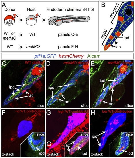
HGF/Met signaling is required in intrapancreatic ducts for pancreatic tail morphogenesis. (A) Schematic of cell transplantation experiments. WT or metMO-injected donor cells were transplanted into WT hosts (top), and WT donor cells were transplanted to metMO-injected hosts (bottom); cas mRNA injection biased donor cells toward endodermal differentiation. (B) Scheme used to identify and quantify contribution of transplanted cells in chimeras. Donor cells are marked by Tg(hs:mCherry) (red), acinar cells are marked by Tg(ptf:GFP)jh1 (blue), and Alcam immunostaining (green) delineates the ducts. (C–E) Single plane confocal images of WT (C) and metMO (D,E) donor endoderm transplanted into WT hosts: WT donor cells contributed to both intrapancreatic ductal (ipd) and acinar (ac) compartments, but metMO donor cells were excluded from the ducts. (F–H) WTkmetMO hosts: With no WT contribution to the pancreas, metMO morphants show donut-like phenotypes (E); with high contribution of WT donor cells, the pancreatic tail growth is rescued (G), and the tip of the tail is almost entirely comprised of WT donor cells (inset of single confocal slice); with moderate contribution of WT donor cells, the pancreatic tail morphology can be rescued with only WT ductal cells (H), and no WT acinar cells are observed in the tip (inset of single confocal slice). |