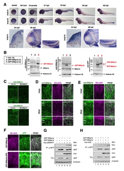
Expression of zebrafish BNip1 mRNA, subcellular localization of zebrafish BNip1 protein, and the in vitro interaction between zebrafish BNip1, !-SNAP and Bcl2 proteins, related to Figure 3
(A) Expression of mRNAs for BNip1a and BNip1b in whole-mount embryos (upper panels). Both mRNAs are expressed ubiquitously at the shield and tail bud stages, and prominently expressed in the head including the retina after 24 hpf. The lower panels indicate retinal sections at 24 and 48 hpf. A higher magnification of photoreceptor cell layer in the ventral retina is indicated. (B) Western blotting of zebrafish 7 hpf non-injection embryos, embryos injected with mRNA of GFP-tagged BNip1a and GFP-tagged BNip1b using anti-BNip1a, anti-BNip1b, and anti-GFP antibodies. Black and red arrows indicate bands corresponding to BNip1a/BNip1b (26 kDa) and GFP-tagged BNip1a/BNip1b, respectively. Anti-BNip1a and anti-BNip1b antibodies specifically detect GFP-tagged BNip1a and BNip1b, respectively. Anti-GFP antibody detected both GFP-tagged BNip1a and GFP-tagged BNip1b. Histone H3 is a loading control detected on the same membrane. (C) Labeling of 48 hpf wild-type and bnip1a and bnipb morphant retinas with anti-BNip1a and anti-BNip1b antibodies, respectively. Antibody signals disappear in the morphants, confirming the specificity of anti-BNip1 antibodies for immunohistochemistry. (D–E) Labeling of 24 and 48 hpf wild-type retinas with anti-BNip1a (D) and anti-BNip1b (E) antibodies (green). ER-mKO (magenta) was used to visualize the ER. The ER is localized in the peri-nuclear region. BNip1a and BNip1b expression patterns overlapped with that of ER-mKO, although the expression pattern of BNip1b is broader than that of BNip1a. Both proteins are detected in the ONL at 48 hpf. (F) Confocal scanning of 24 hpf wild-type retinas expressing ER-mKO as well as GFP-tagged BNip1b (top panels) and GFP-tagged BNip1b-!TM (bottom panels), the latter of which lacks the transmembrane domain (TM). GFP-tagged BNip1b (green) is localized to mesh-like subcellular structures surrounding the nucleus and its distribution overlaps with that of ER-mKO (magenta), suggesting that GFP-tagged BNip1b is enriched in the ER. Conversely, GFP-tagged BNip1b-ΔTM does not overlap with ER-mKO, but rather, is uniformly located outside the nucleus, suggesting that TM is required for ER-localization of BNip1. (G, H) Interaction between zebrafish β-SNAP1 and zebrafish BNip1 proteins (G), and between zebrafish Bcl2 and zebrafish BNip1 proteins (H). Detection of these interactions in HEK293 cells by co-immunoprecipitation. IP: antibody used for immunoprecipitation; Blot: antibodies used for western blotting. TCL: total cell lysates.
|

