
Fig. 3
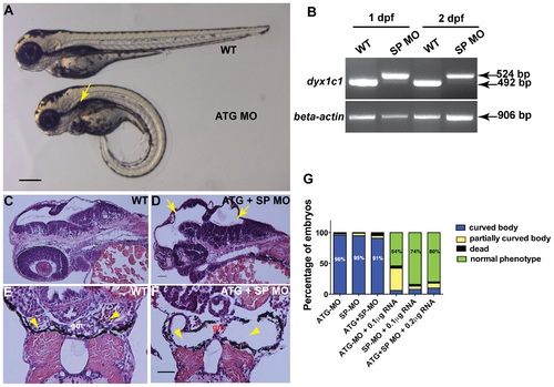
Knockdown of dyx1c1 showed typical cilia phenotypes. Injection of ATGMO or SPMO at 200 µM concentration produced ventrally curved body axis, hydrocephalus and kidney cysts (A). Arrow in panel A denotes kidney cyst in ATG morphant. RT-PCR showed aberrant splice transcripts in SPMO injected embryos at 1 and 2 dpf (B). Histological sections of 2 day old embryos injected with both ATGMO and SPMO (100 µM each) showed hydrocephalus (D; yellow arrows) compared to normal size brain ventricles in WT (C). Transverse histological sections across the pronephros at 3.5 dpf showed normal pronephros in WT embryos (E). Section of dyx1c1 morphant (ATGMO+SPMO) showed severe pronephric distention and a thin glomerulus in the center (F). Yellow arrowheads in panel E and F point out normal pronephros in WT and dilated pronephros in morphant embryo, respectively. Quantitative analysis of the rescue of dyx1c1 morphant phenotype to WT phenotype with different combinations of MOs and dyx1c1 mRNA (G). Scale bars indicate 100 µm. Abbreviation: gm, glomerulus. |
| Fish: | |
|---|---|
| Knockdown Reagents: | |
| Observed In: | |
| Stage Range: | Long-pec to Protruding-mouth |