
Fig. 8
- ID
- ZDB-FIG-130619-29
- Publication
- Nair et al., 2013 - The chromosomal passenger protein birc5b organizes microfilaments and germ plasm in the zebrafish embryo
- Other Figures
- All Figure Page
- Back to All Figure Page
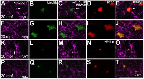
Birc5b localizes to cortical microtubule tips and co-localizes with F-actin and germ plasm RNPs. High magnifications of animal views of blastodisc cortex. In wild-type embryos, Birc5b (B, L) localizes to monoaster microtubule tips (A, C, K, M, arrows) and polymerizing F-actin (D, E). In motley/birc5b mutants (F–J) Birc5b co-localizes with polymerizing F-actin (I, J), but as tips of the microtubules are not seen at the cortex (F), co-localization to the microtubule tip is not observed. Birc5b at the microtubule tips (K, M arrow) co-localizes with GP RNPs in wild-type embryos (N, O). In motley/birc5b mutants (P–T), microtubule tips are not seen at the cortex (P) but Birc5b continues to localize with GP RNPs (S, T). |
| Gene: | |
|---|---|
| Antibodies: | |
| Fish: | |
| Anatomical Terms: | |
| Stage: | 1-cell |