
Fig. S2
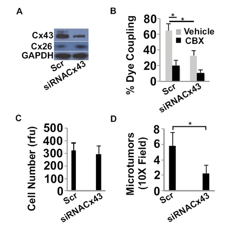
siRNA depletion of Cx43 inhibits GJ communication, and microtumor formation in the brain. (A) 4T-1 cells were treated with a scrambled control RNA (Scr) or siRNA to Cx43 (siRNAcx43) and then western blotted for the indicated proteins. (B) The indicated tumor cells were prelabeled with calcein orange dye and then added to a monolayer of EA.hy926 endothelial cells in the presence of the GJ inhibitor CBX (10 μM) or its vehicle (PBS). Dye transfer from tumor cells to endothelial cells was observed live by epifluorescence microscopy after 30 minutes of co-culture. The number of adherent cells that transferred dye to the adjacent endothelium was determined and represented as percent of total number of tumor cells counted. * = P values < 0.05 (C) The indicated tumor cells were cultured in vitro and examine for cell growth for 3 days in the presence of CBX or its vehicle using the CyQUANT assay. rfu = relative florescence units. (D) 4T-1scr or 4T-1siRNAcx43 cells expressing GFP were injected into the chicken circulation and allowed to form microtumors in the brain. The number of microtumor lesions per 10X field in the brain was determined by confocal microscopy 5 days after injection. * = P values < 0.05. |