
Fig. 5
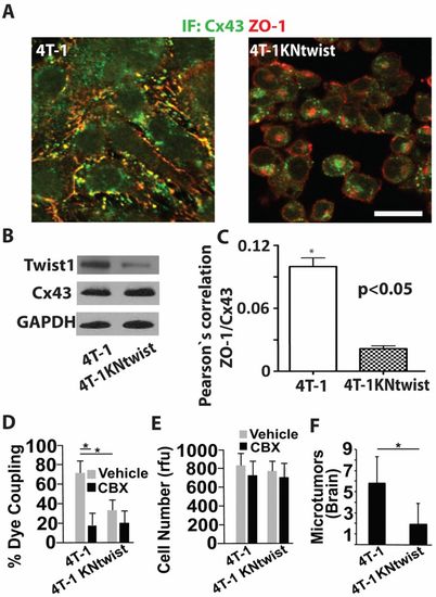
Twist inhibition in metastatic breast cancer cells reduces Cx43-mediated GJ communication and inhibits brain microtumor formation. (A) Representative images show 3D reconstructions of confocal z-stacks (60×, top views) of 4T-1 expressing the empty lentiviral vector or 4T-1 cells expressing lentiviral vectors encoding shRNAs to twist (4T-1KNtwist). Cells were co-immunostained for Cx43 and ZO-1 and imaged as for Fig.4A. Note that in cells with twist depleted, Cx43 is mostly intracellular, whereas ZO-1 is localized to the membrane. Scale bar: 20μm. (B) Western blots of indicated proteins in 4T-1 and 4T-1KNtwist cells. Note that loss of twist expression inhibits membrane localization of Cx43 as shown in A, but not Cx43 total protein levels. 4T-1KNtwist cells show a 71% decrease in Twist1 expression compared to 4T-1 cells, as measured by densitometry. (C) Pearson′s correlation analyses of Cx43 and ZO-1 colocalization in 4T-1 and 4T-1KNtwist cells immunostained as for A. (D) 4T-1 and 4T-1KNtwist cells in the presence of CBX (10μM) or vehicle were examined for GJ-mediated calcein orange dye transfer to the cultured endothelium as described for Fig.2B. (E) 4T-1 and 4T-1KNtwist cell growth in the presence of CBX (10μM) or vehicle was determined as for Fig.2C. (F) The number of 4T-1 and 4T-1KNtwist microtumors was assessed after 7 days in the chicken brain as described for Fig.3D. Data indicate means + s.e.m. *P<0.05. |