
Fig. S2
- ID
- ZDB-FIG-120810-40
- Publication
- Feng et al., 2012 - Live Imaging of Tumor Initiation in Zebrafish Larvae Reveals a Trophic Role for Leukocyte-Derived PGE(2)
- Other Figures
- All Figure Page
- Back to All Figure Page
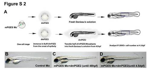
Schematic to illustrate mPGES morpholino experiment + larval images to show the normal development of treated larvae (related to Figure 1). (A) Schematic outline of our procedure for morpholino knockdown of mPGES expression in zebrafish larvae. In both treated groups the medium was supplemented with stable PGE2 (dmPGE2) to compensate for the PGE2 required for normal development up to 42hpf; subsequently, dmPGE2 was removed from the PGE2 suppressed group. (B-D) Wide field images of the morphologies of Control morphant (B), mPGES Morphant with dmPGE2 rescued until 48hpf (C), and mPGES Morphant larvae with dmPGE2 rescued until 4.5dpf (D). |