
Fig. 5
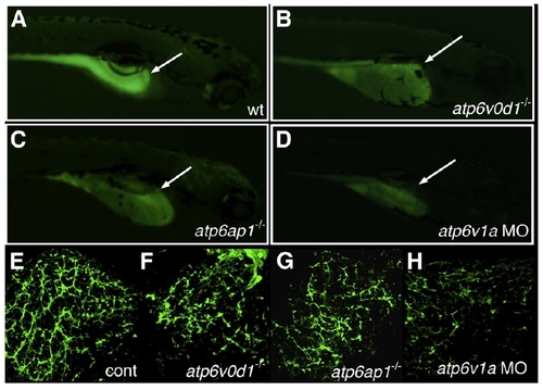
Intrahepatic biliary defects in other V-H+-ATPase mutants. (A–D) Right lateral views of 5 dpf larvae after administration of the fluorescent lipid reporter PED-6. (A) Control larva demonstrates normal gallbladder intensity (white arrow). (B) 5 dpf atp6v0d1-/- mutant, (C) 5 dpf atp6ap1-/- mutant, and (D) 5 dpf atp6v1a morpholino (MO)-injected larvae all demonstrate a decrease or lack of gallbladder fluorescence (white arrows). (E–H) Confocal projections of cytokeratin immunostaining of 5 dpf liver from control (E), atp6v0d1-/-, atp6ap1-/-, and atp6v1a MO-injected larvae. The abnormal cytokeratin pattern in (F–H) has similarities to the pattern noted in pn, with fewer ducts overall and focal areas of ectatic ducts. |
| Fish: | |
|---|---|
| Knockdown Reagents: | |
| Observed In: | |
| Stage: | Day 5 |
Reprinted from Developmental Biology, 365(2), Eauclaire, S.F., Cui, S., Ma, L., Matous, J., Marlow, F.L., Gupta, T., Burgess, H.A., Abrams, E.W., Kapp, L.D., Granato, M., Mullins, M.C., and Matthews, R.P., Mutations in vacuolar H(+)-ATPase subunits lead to biliary developmental defects in zebrafish, 434-444, Copyright (2012) with permission from Elsevier. Full text @ Dev. Biol.