
Fig. 2
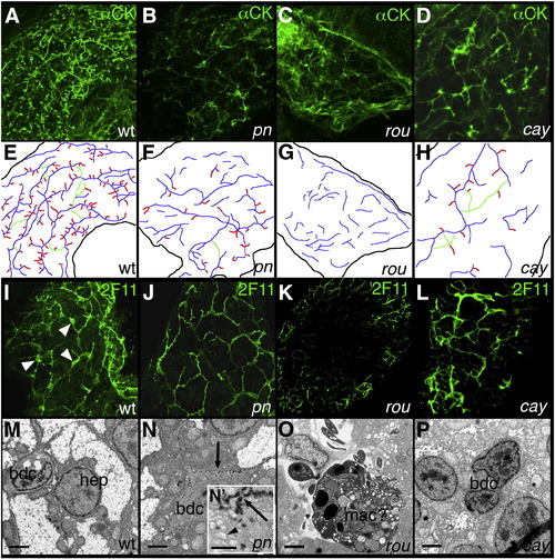
Intrahepatic biliary defects in novel mutants. (A–D) Confocal projections of whole-mount cytokeratin immunostaining of livers from 5 dpf wild-type (A, wt), pekin (B, pn), rouen (C, rou), and cayuga (D, cay) larvae, schematized in (E–H). The typical wt pattern is depicted in (A) and schematized in (E), demonstrating a complex latticework. In contrast, the staining pattern of pn (B, F) demonstrates shorter ducts with fewer interconnections. The staining pattern of rou (C, G) demonstrates an absence of recognizable ducts. The cytokeratin staining pattern of cay (D, H) demonstrates short non-interconnected ducts, with some terminal ductules. (I–L) Confocal projections of whole-mount 2F11 immunostaining of livers from 5 dpf wt (I), pn (J), rou (K), and cay (L) larvae. 2F11 stains bile duct cell bodies (white arrowheads) and proximal ducts, as demonstrated in wt (I). The 2F11 pattern of pn demonstrates fewer ducts, with smaller bile duct cells. There are no apparent bile duct cells in 2F11 stainings of rou (K), and cay (L) is similar to wt. (M–P) Electron micrographs of livers from 5 dpf wt (M), pn (N), rou (O), and cay (O) larvae. In (M), the wt hepatocyte (hep) and bile duct cell (bdc) are depicted. In contrast, the bdc in pn (N) contains an accumulation of electron dense material (black arrow) at the periphery of the cell. The inset (N2) demonstrates that these bodies have a striped appearance, and the dilated vesicles (black arrowhead) are also apparent. The cell in rou (O) is most likely a macrophage (mac), in the space normally occupied by bile duct cells. In contrast, the bdc of cay (P) appears close to normal, but with nuclei that are modestly enlarged relative to the cytoplasm when compared to wt (M), and with an appearance more closely resembling the hep nucleus. Scale bars in (M–P) 500 nm, N2 also 500 nm. |
| Antibody: | |
|---|---|
| Fish: | |
| Anatomical Term: | |
| Stage: | Day 5 |
| Fish: | |
|---|---|
| Observed In: | |
| Stage: | Day 5 |
Reprinted from Developmental Biology, 365(2), Eauclaire, S.F., Cui, S., Ma, L., Matous, J., Marlow, F.L., Gupta, T., Burgess, H.A., Abrams, E.W., Kapp, L.D., Granato, M., Mullins, M.C., and Matthews, R.P., Mutations in vacuolar H(+)-ATPase subunits lead to biliary developmental defects in zebrafish, 434-444, Copyright (2012) with permission from Elsevier. Full text @ Dev. Biol.