
Fig. 3
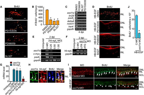
HB-EGF and Retinal Injury Stimulate MG Dedifferentiation via an EGFR/MAPK/Ascl1a-Signaling Cascade (A) EGFR inhibition (PD153035) or MAPK inhibition (PD98059, SL327) reduces the number of proliferating progenitors generated following retinal injury. Asterisks identify the injury sites. (B) Quantification of (A). **p < 0.01. (C) RT-PCR shows that EGFR or MAPK inhibition blocks induction of genes associated with MG dedifferentiation. (D) EGFR or MAPK inhibition reduces the number of proliferating progenitors generated following HB-EGF treatment of the uninjured retina. (E) HB-EGFa knockdown suppresses induction of regeneration-associated genes in the injured retina. [hb-egf MO] is in mM. C-MO is 0.5 mM. (F) Ascl1a knockdown has little effect on hb-egfa induction in the injured retina (MOs are 0.25 mM). (G) Real-time PCR quantification of (E) and (F) normalized to uninjured value. The hb-egf MO is at 0.5 mM, and ascl1a MOs are at 0.25 mM. (H) ascl1a and hb-egfa in situ hybridization assays along with BrdU immunofluorescence show colocalization (arrows) in the injured retina. (I) MO-mediated knockdown of Ascl1a in the presence of HB-EGF suppresses HB-EGF-dependent generation of BrdU+ progenitors. BrdU+ progenitors that do form in the Ascl1a knockdown retina generally lack lissamine-labeled MO (arrowheads). Control MO does not block the generation of BrdU+ progenitors (arrows). (J) Quantification of BrdU+ cells in (I). *p < 0.01. Error bars represent SD. All scale bars, 50 μm. ONL, outer nuclear layer; INL, inner nuclear layer; GCL, ganglion cell layer. See also Figure S3. |
| Genes: | |
|---|---|
| Fish: | |
| Condition: | |
| Knockdown Reagents: | |
| Anatomical Term: | |
| Stage: | Adult |
Reprinted from Developmental Cell, 22(2), Wan, J., Ramachandran, R., and Goldman, D., HB-EGF Is Necessary and Sufficient for Müller Glia Dedifferentiation and Retina Regeneration, 334-347, Copyright (2012) with permission from Elsevier. Full text @ Dev. Cell