
Fig. 1
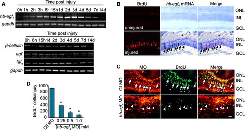
Injury-Dependent hb-egfa Induction Is Necessary for MG Dedifferentiation and Proliferation (A) RT-PCR showing the temporal expression pattern of genes encoding EGFR ligands in the uninjured and injured retina. (B) In situ hybridization and BrdU immunofluorescence show that hb-egfa is induced in proliferating progenitors in the injured retina (arrows) (* marks the injury site). (C) MO-mediated HB-EGFa knockdown reduces the number of proliferating MG-derived progenitors in the injured retina at 4 dpi. Arrows identify proliferating cells in the control MO-treated retina, whereas arrowheads identify proliferating cells in the HB-EGFa knockdown retina (note that these latter cells lack lissamine-labeled MO). (D) Quantification of (C) at different hb-egfa MO concentrations. *p < 0.01. Error bars represent SD. Scale bars, 50 μm. ONL, outer nuclear layer; INL, inner nuclear layer; GCL, ganglion cell layer. |
| Genes: | |
|---|---|
| Fish: | |
| Condition: | |
| Anatomical Terms: | |
| Stage: | Adult |
| Fish: | |
|---|---|
| Condition: | |
| Knockdown Reagent: | |
| Observed In: | |
| Stage: | Adult |
Reprinted from Developmental Cell, 22(2), Wan, J., Ramachandran, R., and Goldman, D., HB-EGF Is Necessary and Sufficient for Müller Glia Dedifferentiation and Retina Regeneration, 334-347, Copyright (2012) with permission from Elsevier. Full text @ Dev. Cell